
Screening With Breast Implants: How Three Imaging Tests Fit Together
Implants change how breast imaging is done, not whether it should be done — and each modality answers a different question.
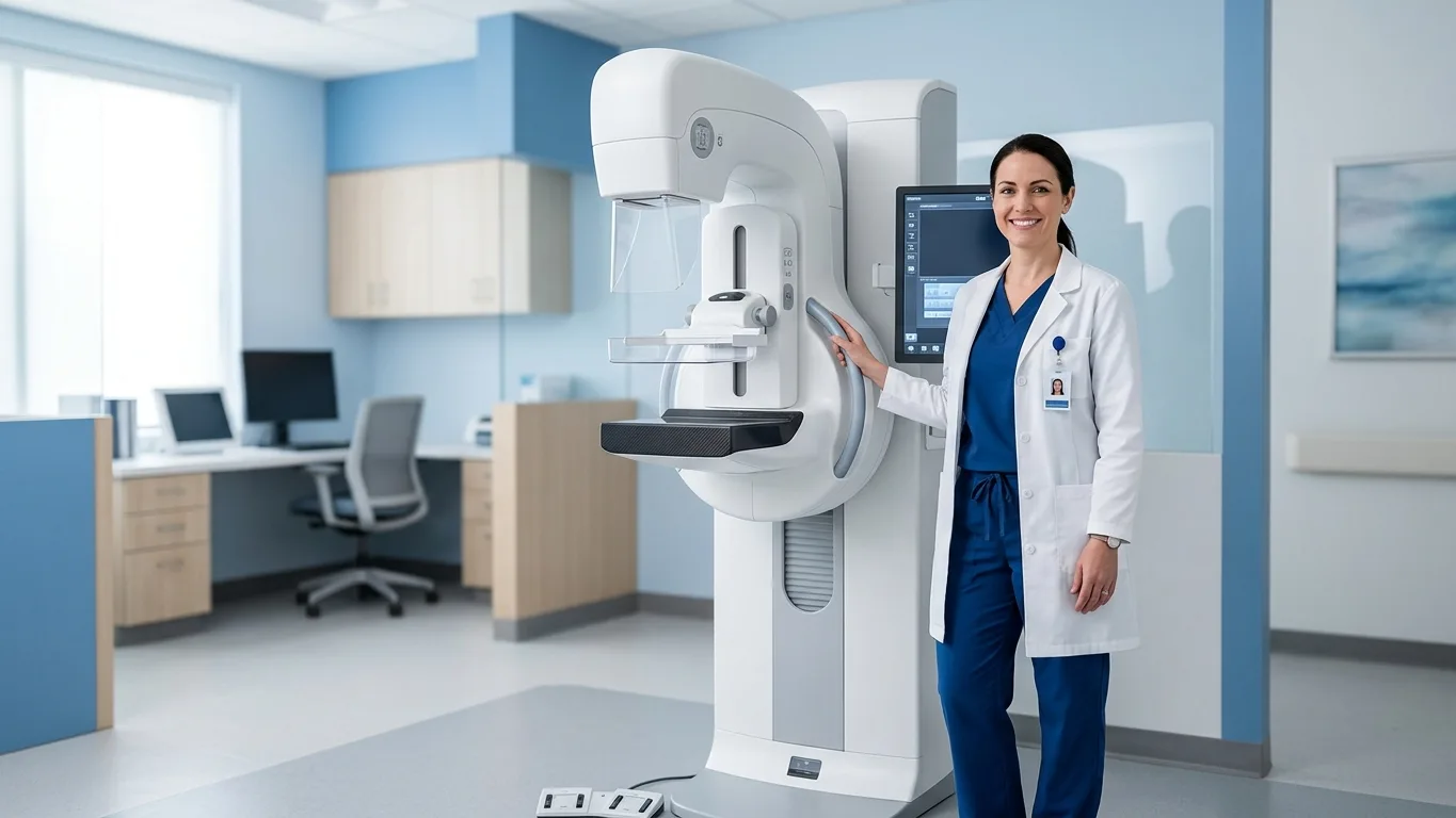
A common misconception holds that breast implants make routine screening pointless or unsafe. In practice, people with implants follow the same age-based screening principles as everyone else — the imaging simply requires technique adjustments and, sometimes, additional tools.
Mammography, ultrasound and MRI each see different things: calcifications, tissue architecture and the implant shell itself. Used together on a sensible schedule, they cover both breast health and device condition — two jobs a single test rarely does alone.
Mammography still works — with displacement views
Standard mammography compresses the breast to image its tissue, and an implant can hide part of that tissue from view. Radiology teams solve much of this with implant-displacement views, often called Eklund views, in which the implant is gently pushed back toward the chest wall so more natural tissue enters the frame. Telling the imaging center about implants in advance lets staff plan these extra views.
Compression is applied carefully, and modern implants are built to tolerate properly performed mammography; worries about routine imaging damaging a device are largely historical. What matters more is consistency — skipping mammograms because of implants removes the one test designed to catch early calcifications.
Where ultrasound and MRI take over
Ultrasound adds a real-time look at tissue near the implant and helps characterize lumps that mammography flags, without radiation. It is also a practical first tool when a patient or physician feels a change on exam, since it distinguishes fluid-filled from solid findings.
MRI is the most sensitive test for the implant shell itself, particularly for silent shell changes that produce no symptoms, and regulators recommend periodic imaging of silicone implants for exactly this reason. Whatever the modality, imaging findings require medical interpretation — a scan is a data point a physician reads in context, never a verdict on its own.
Building a routine that lasts
A workable breast implant screening plan usually layers the tests: age-appropriate mammography on the standard schedule, ultrasound when a question needs answering, and periodic MRI for silicone devices per current guidance. The right cadence differs with age, family history, breast density and implant type — individual variation a physician weighs when setting intervals.
Routine checks catch changes early, when options are widest. No pathway is flawless: callbacks, false alarms and — with contrast studies — a small possibility of side effects all exist, and the balance differs person to person. Discussing the schedule with a board-certified specialist, and revisiting it after any breast surgery once the recovery period has passed, keeps screening aligned with the body it is watching.
Before your next screening visit
- Tell the imaging center you have implants when booking, so displacement views are planned.
- Bring implant details if you have them — type, year of surgery and any device card.
- Note any changes in shape, firmness or sensation since your last visit.
- Ask how mammography, ultrasound and MRI will be sequenced in your case.
- Keep prior images accessible; comparison is half the value of screening.
- Consult a board-certified specialist to interpret results — imaging findings need medical judgment.
MediIndex articles are for general information only and are not medical advice, diagnosis, or advertising. Outcomes vary by individual — consult a board-certified specialist for personal decisions.