Mammary Tumors in Dogs: Why Early Spaying Changes the Odds
Veterinarians explain why mammary lumps are so common in unspayed older dogs, and why surgery with a pathology report remains the standard of care.
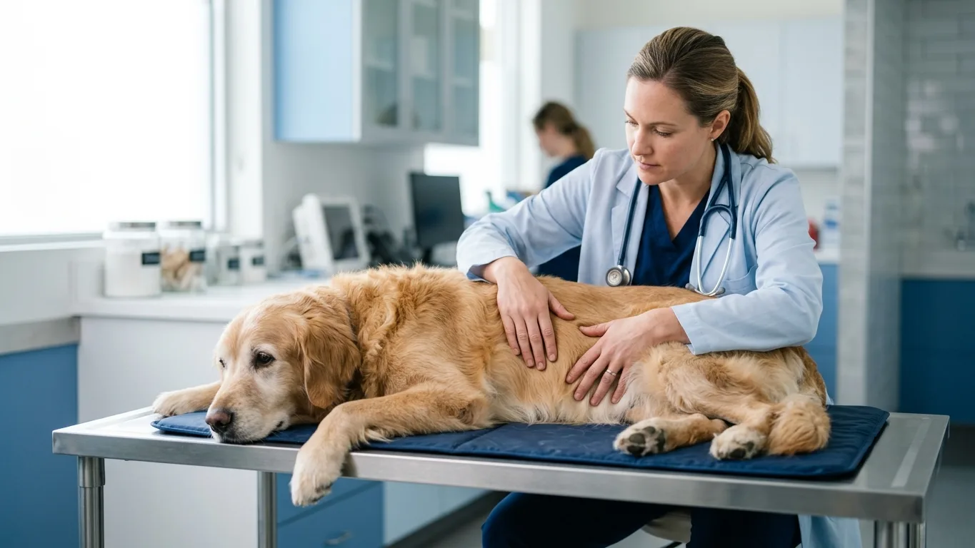
A firm lump along a dog’s belly is one of the findings veterinarians take most seriously in older, unspayed females. Mammary tumors are among the most common tumors in this group, and looks alone cannot tell a harmless growth from a dangerous one. That uncertainty is exactly why every new lump deserves an in-person veterinary exam rather than a wait-and-see approach at home.
The disease is closely tied to hormone exposure. Dogs spayed before their first or second heat cycle face a far lower lifetime risk, while intact females who reach middle age carry the highest burden. For a dog that already has a lump, the path is well established: examination, staging tests, surgical removal and a histopathology report that names what the mass actually is.
Why hormones set the stage
Each heat cycle bathes mammary tissue in estrogen and progesterone, and repeated cycles appear to prime that tissue for tumor formation years later. This is why spay timing matters so much: removing the ovaries early in life removes most of that cumulative exposure. Veterinary groups still weigh spay timing against breed, size and orthopedic considerations, so the right schedule is a case-by-case conversation with a veterinarian.
Risk is not uniform, either. Age, breed and each dog’s own biology all shift the picture, and some spayed dogs still develop mammary tumors while some intact dogs never do. Individual variation is real, which is why routine belly checks and regular veterinary visits matter for every female dog, whatever her spay status.
Surgery plus histopathology: the standard path
Needle sampling of a mammary mass often cannot distinguish benign from malignant disease, so surgical removal is usually both the diagnosis and the treatment. Before surgery, veterinarians typically run bloodwork and chest imaging to check overall health and look for any sign of spread. Those results shape how wide the surgery needs to be.
What happens after the mass comes out is just as important. The tissue goes to a pathology laboratory, where histopathology identifies the tumor type, its grade and whether the surgical margins are clean. That report—not the surgery itself—tells the veterinary team whether monitoring is enough or further treatment should be discussed.
Recovery, risks and the long watch
Recovery from mammary surgery generally takes a couple of weeks of restricted activity while the incision heals, though healing speed varies from dog to dog. As with any surgery under anesthesia, complications are possible—incision swelling, infection or fluid buildup among them—so owners are asked to watch the site daily and report changes promptly. Follow-up rechecks let the veterinary team catch problems early.
Even after a clean report, new lumps can appear in remaining mammary tissue over time. Veterinarians usually recommend monthly at-home belly checks plus scheduled rechecks so anything new is measured, mapped and assessed early. If a new or changing lump turns up, the next step is not the internet—it is a veterinary appointment.
Before your vet visit
- Note when you first felt the lump and whether its size or texture has changed.
- Bring your dog’s spay status and heat-cycle history, as precisely as you know it.
- List all medications and supplements your dog currently takes.
- Track recent appetite, weight and energy changes to share with the veterinarian.
- Photograph or measure the lump so changes can be compared over time.
MediIndex articles are for general information only and are not medical advice, diagnosis, or advertising. Outcomes vary by individual — consult a board-certified specialist for personal decisions.