Gallbladder Mucocele in Dogs: The Silent Problem Ultrasound Finds
A gallbladder mucocele can build for months without obvious signs, which is why veterinarians lean on abdominal ultrasound to catch it before it ruptures.
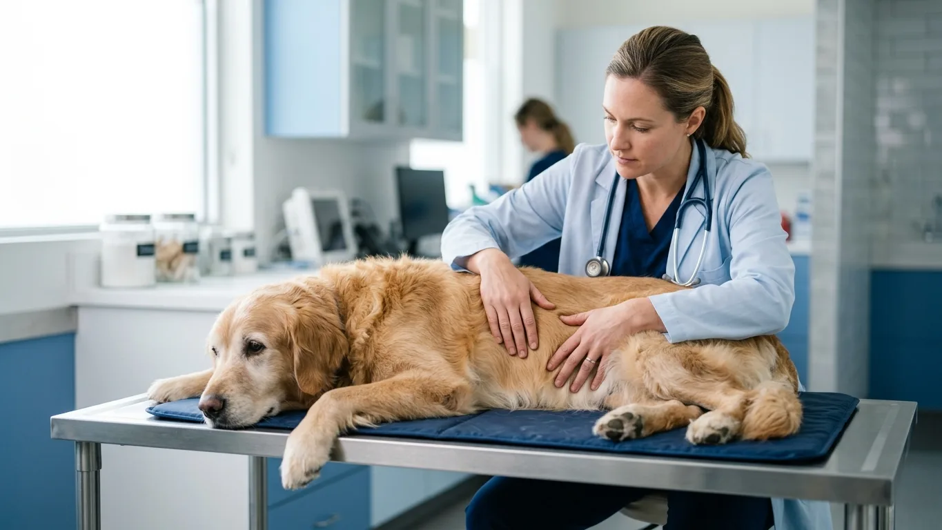
Some of the most dangerous problems in veterinary medicine make almost no noise, and the gallbladder mucocele is a leading example. Bile inside the gallbladder gradually turns into a thick, gelatinous mass that stretches the organ’s wall, often while the dog eats, plays and behaves normally. Many mucoceles are discovered only when an abdominal ultrasound is performed for another reason.
When signs do appear—vomiting, poor appetite, lethargy, belly pain or yellow-tinged gums—the disease may already be advanced, and a ruptured gallbladder can spill bile into the abdomen. Any of those signs is a reason to see a veterinarian promptly rather than wait at home. How fast a mucocele progresses varies widely between individual dogs, which is why veterinarians treat every confirmed case as one to watch closely.
Why the gallbladder goes quiet, then critical
The gallbladder stores bile between meals, and in a mucocele that bile is replaced by immobile, mucus-laden material that cannot drain normally. Pressure builds slowly, the wall weakens, and outflow to the intestine can become blocked. Because the process unfolds over months, the body adapts and outward signs stay subtle until late.
Middle-aged and older dogs are diagnosed most often, and some breeds, including Shetland sheepdogs and cocker spaniels, appear predisposed. Hormonal diseases and abnormal blood-fat levels have also been linked to the condition. Still, individual variation is wide—a predisposed dog may never develop one, while a dog with no known risk factors can.
Ultrasound: how a silent problem gets caught
Abdominal ultrasound is the tool that most often reveals a mucocele, showing the gallbladder filled with organized, non-moving material instead of free-flowing bile. Veterinarians describe a characteristic layered or star-like pattern in advanced cases. Bloodwork frequently adds supporting clues, such as elevated liver and bile-related values.
An ultrasound image is not a verdict on its own—early mucoceles can resemble ordinary thickened bile, and the findings must be read alongside bloodwork and the dog’s history. That interpretation belongs to a veterinarian, not to a screenshot compared against pictures online. Regular imaging rechecks are how mild changes are separated from a mucocele that is progressing.
Surgery, monitoring and the road back
For a mucocele that is advanced or already causing signs, removing the gallbladder—a cholecystectomy—is the most commonly recommended path, while some early, incidental cases are managed with medication and scheduled ultrasound rechecks. Which route fits a given dog depends on the imaging picture, bloodwork and overall health, and it is a decision made with a veterinarian in person.
Surgery on a fragile, bile-filled organ carries real risks, and complications such as bile leakage, pancreatitis or infection are possible, particularly if the gallbladder has already ruptured. Most dogs need a recovery period of roughly two weeks of restricted activity with follow-up bloodwork, though healing speed differs from dog to dog. Owners are asked to watch appetite, energy and gum color closely during that window and report changes to the clinic without delay.
Before your vet visit
- Write down any vomiting, appetite dips or low-energy days from the past few weeks, with dates.
- Bring prior ultrasound reports or bloodwork results if your dog has them.
- List current medications, supplements and diet, including recent changes.
- Note any hormonal conditions your dog has been diagnosed with.
- Check your dog’s gums in daylight and mention any yellow tint to the veterinarian.
MediIndex articles are for general information only and are not medical advice, diagnosis, or advertising. Outcomes vary by individual — consult a board-certified specialist for personal decisions.